Surgeons may soon be able to localize critical regions in tissues and organs during a surgical operation thanks to a new Purdue University biosensor that can be printed in 3D using an automated printing system.
Chi Hwan Lee, Leslie A. Geddes Assistant Professor of Biomedical Engineering in the Weldon School of Biomedical Engineering and assistant professor of mechanical engineering, created the biosensor, which allows for simultaneous recording and imaging of tissues and organs during a surgical operation.
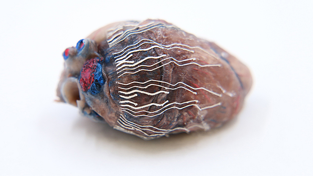
“Simultaneous recording and imaging could be useful during heart surgery in localizing critical regions and guiding surgical interventions such as a procedure for restoring normal heart rhythms,” Lee said.
Traditional methods to simultaneously record and image tissues and organs have proven difficult because other sensors used for recording typically interrupt the imaging process.
“To this end, we have developed an ultra-soft, thin and stretchable biosensor that is capable of seamlessly interfacing with the curvilinear surface of organs; for example the heart, even under large mechanical deformations, for example cardiac cycles,” Lee said.
“This unique feature enables the simultaneous recording and imaging, which allows us to accurately indicate the origin of disease conditions: in this example, real-time observations on the propagation of myocardial infarction in 3D.”
By using soft bio-inks during the rapid prototyping of a custom-fit design, biosensors fit a variety of sizes and shapes of an organ. The bio-inks are softer than tissue, stretch without experiencing sensor degradation and have reliable natural adhesion to the wet surface of organs without needing additional adhesives.
Scientist Kwan-Soo Lee’s research group in Los Alamos National Laboratory is responsible for the formulation and synthesis of the bio-inks. A number of prototype biosensors using different shapes, sizes and configurations have been produced.
Craig Goergen, the Leslie A. Geddes Associate Professor of Biomedical Engineering in Purdue’s Weldon School of Biomedical Engineering, and his laboratory group have tested the prototypes in mice and pigs in vivo.
“Professor Goergen and his team were successfully able to identify the exact location of myocardial infarctions over time using the prototype biosensors,” Lee said.
“In addition to these tests, they also evaluated the biocompatibility and anti-biofouling properties of the biosensors, as well as the effects of the biosensors on cardiac function. They have shown no significant adverse effects.”
The Purdue Research Foundation Office of Technology Commercialization has filed a patent application on Lee’s biosensor. Other steps taken to develop the sensor include exploring further applications of the bio-inks into various printable biosensors with a tailored design to fit other organs such as the stomach, which requires even higher stretchability than the heart.


